26.01.2023
Whilst ultrasound technology continues to evolve and improvements are made in image resolution and the reduction of artefacts, some anatomic areas remain a challenge to the achievement of diagnostic quality images.
One example is the ultrasound examination of superficial structures. Areas close to the transducer contact point on the body can suffer reduced image quality for a combination of reasons:
Some of the factors above can be improved by careful patient preparation, appropriate adjustment of the ultrasound system controls, and the use of higher frequency linear transducers (Figure 1.). They can also be improved through the use of an ultrasound standoff pad.
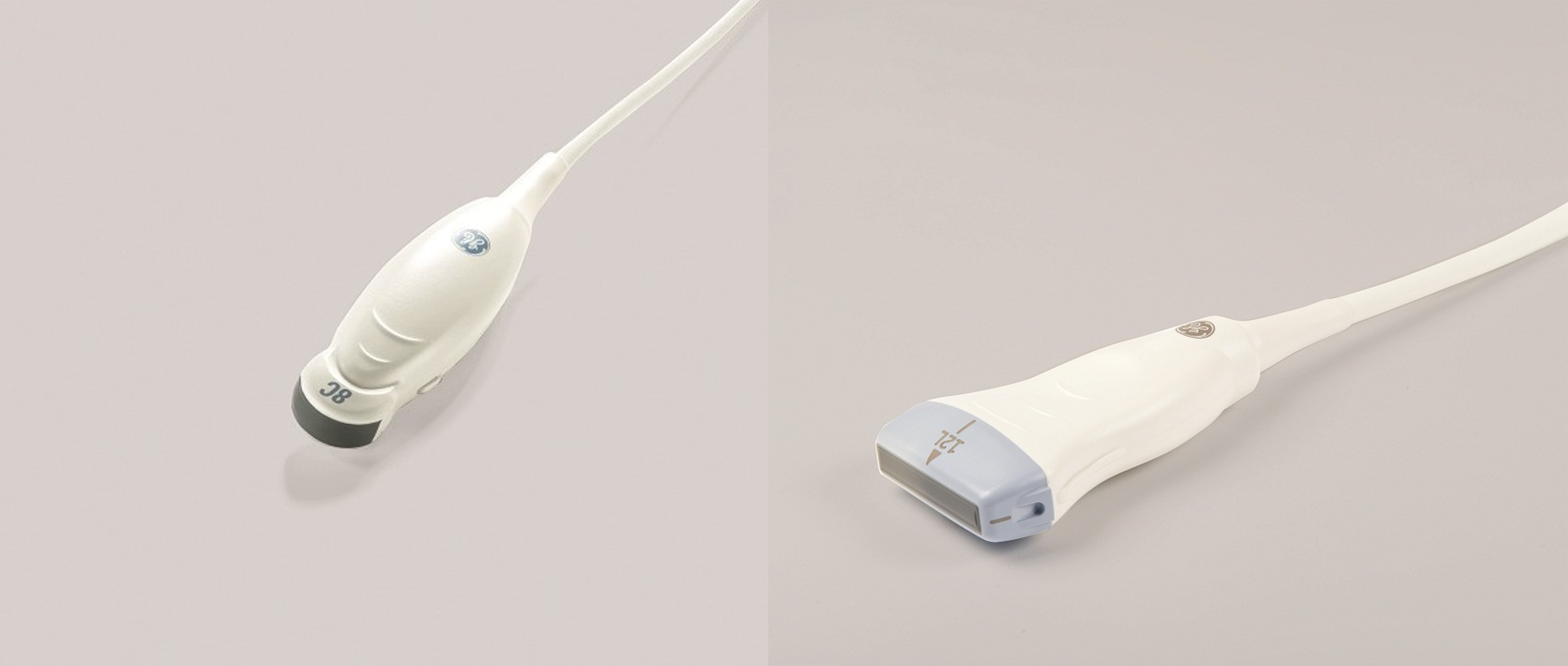
Figure 1. Two different transducer designs. A microconvex transducer design with a curved surface is shown on the left. On the right is a linear design with a flat, rectangular surface.
Standoff pads are typically a flexible, acoustically inert material that is placed between the transducer surface and the patient’s skin. They cause minimal attenuation of ultrasound and typically appear anechoic or black on the ultrasound image.
The use of a standoff pad alleviates many of the reasons for reduced image quality when examining superficial structures and their use is essential to achieving the diagnostic images of some anatomic areas.
Most modern standoff pads are made from silicone or similar polymers and are designed in a variety of shapes from simple sheets or discs, through to models that fit over specific transducer designs (Figure 2.).
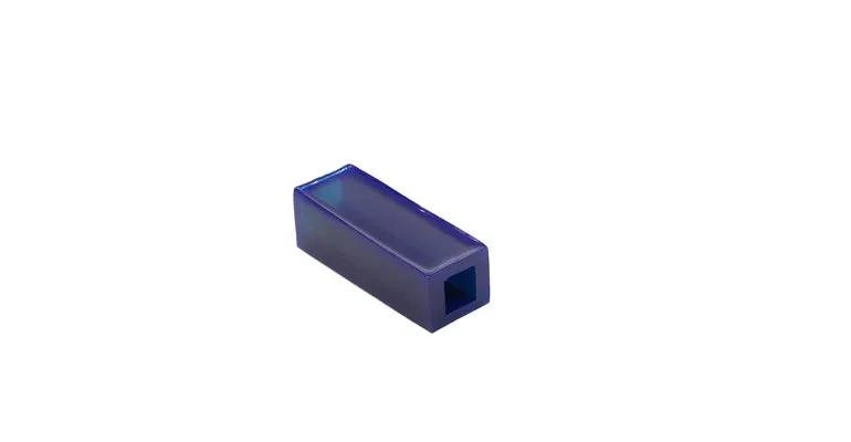
Figure 2. A modern polymer standoff designed to fit over a linear transducer.
Standoff pads can also be made using examination gloves with water or coupling gel. These inexpensive designs can be useful where standoff pads are only needed sporadically. However, it can be difficult to avoid bubbles of air when filling the glove, and any air present will cause artefacts.
In veterinary medicine there are many applications for standoff pads;
A good example is in the examination of the superficial digital flexor tendon (SDFT). By using a standoff, the palmar aspect of the tendon and overlying skin can be more accurately evaluated (Figures 3 and 4).
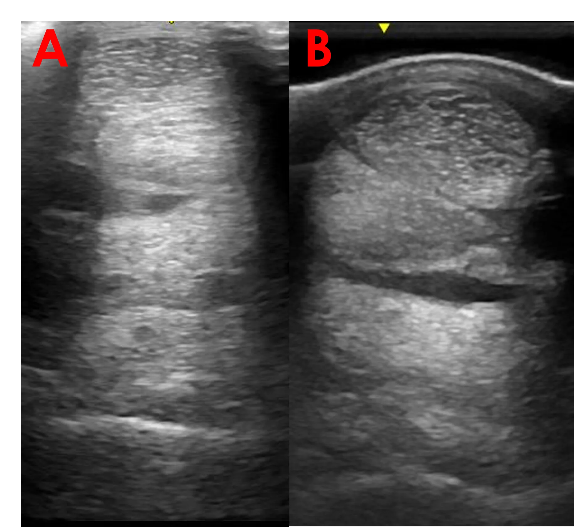
Figure 3. Two transverse plane images of the proximal palmar metacarpal region in a horse. In Image A, a standoff is not used. In Image B, the use of a standoff allows a more accurate evaluation of the palmar SDFT.
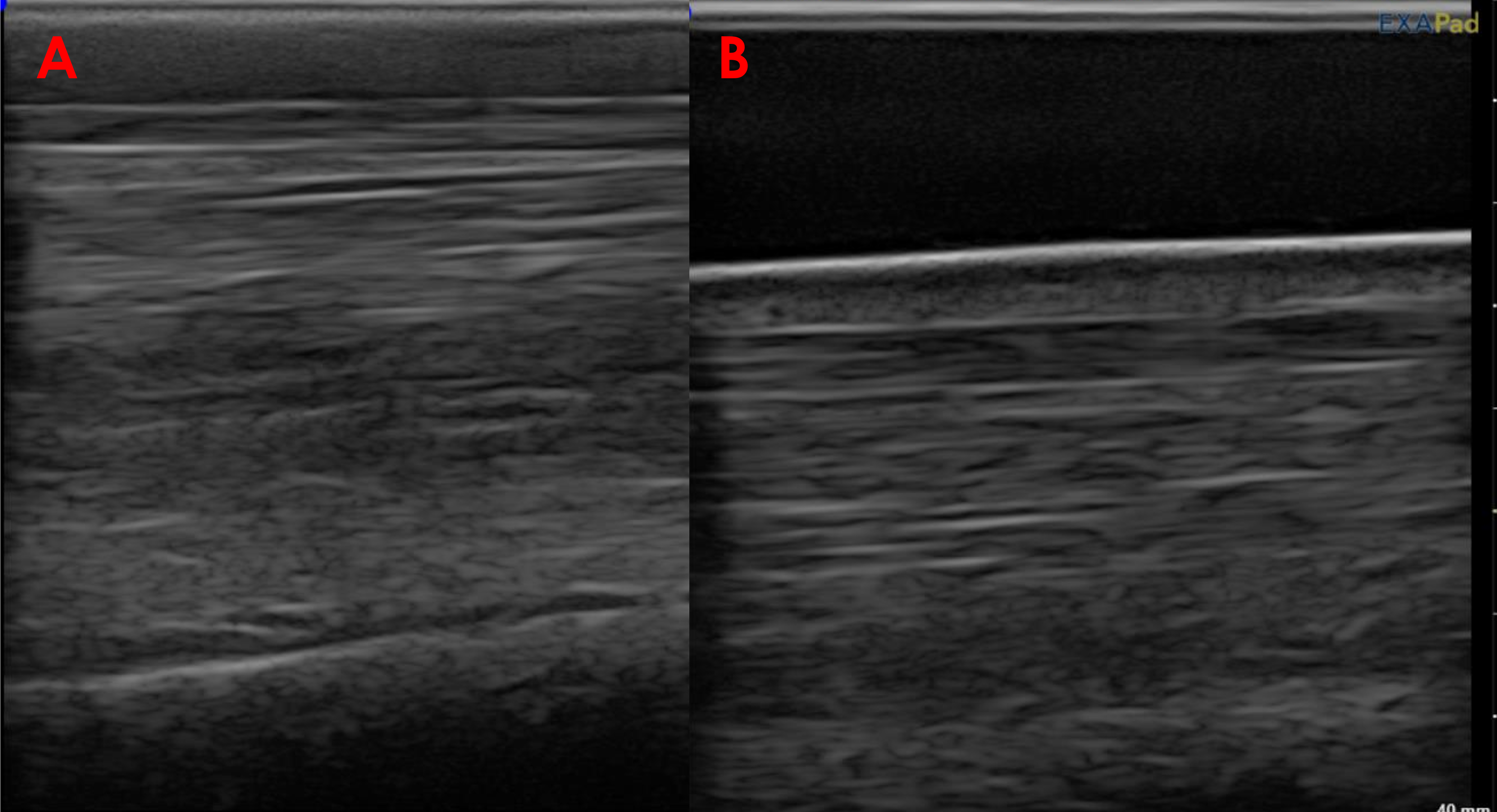
Figure 4. Two longitudinal plane images of the proximal palmar metacarpal region in a horse. In Image A, a standoff is not used. In Image B, the use of a standoff allows a more accurate evaluation of the palmar SDFT and overlying skin.
Despite some of the difficulties of imaging superficial structures, by considering the use of a standoff along with appropriate transducer choice and patient preparation, diagnostic quality ultrasound images can be achieved.